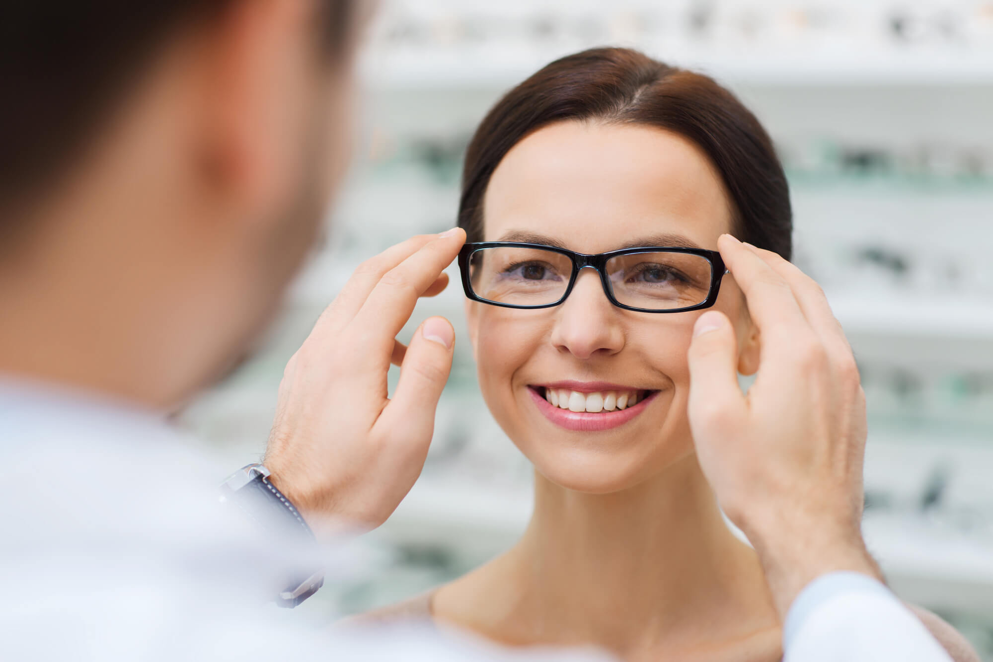
Routine Eye Care
From updating your vision prescription to looking for more serious health issues, annual eye exams are an important part of maintaining your #VisionForLife
Book an appointment
Our Patient-Centric Approach to Treatment
Georgia Ophthalmologists believes to be considered as an individual is an essential part of each patient’s #VisionForLife..
Why do you need an annual eye exam?
A thorough eye exam is important to maintaining overall health.
-
UPDATE PRESCRIPTIONS
Your eyes can change from year to year, and glasses and contact lenses prescriptions should be checked annually.
-
SENIOR EYE CARE
Seniors should be checked annually for signs of cataracts, macular degeneration, and/or glaucoma.
-
DIABETIC EYE CARE
It is recommended that individuals with diabetes undergo annual eye exams to check for any signs of swelling or bleeding in the retina.
-
PEDIATRIC EYE CARE
To ensure that a child’s eyesight is developing normally, it’s recommended that they receive a comprehensive eye exam at least once every two years.
What results are possible?
Our comprehensive eye exams ensure healthy vision by detecting issues early and providing personalized recommendations for treatments. Prevent vision loss and maintain optimal eye health with regular visits.
Do you need an eye exam if you have good vision?
It’s important to have regular eye exams, even is you don’t require corrective lenses, as they can detect early signs of issues such as refractive errors (like farsightedness, nearsightedness, or astigmatism) or conditions like glaucoma.

The Georgia Ophthalmologists Approach
Promoting regular eye exams is our way of ensuring our patients’ #VisionForLife.


Patients Say
Comprehensive Annual Eye Exams
Routine eye examinations are an important part of your #VisionForLife
At Georgia Ophthalmologists, we offer comprehensive annual eye examinations for all ages. During your examination, the doctor will thoroughly evaluate the total health of your eyes, which includes vision screening, pressure reading, dry eye evaluation, dilation which allows the doctor to examine the back of your eye, and refraction which determines your glasses or contact lens prescription.
Most people will seek medical attention for sore or painful eyes, changes in vision, or for an injury, but do not otherwise feel eye examinations are an important part of routine medical care unless a problem occurs. Routine eye exams are a very important as many blinding eye diseases, such as cataracts, glaucoma, macular degeneration and diabetic eye disease, have little or no warning signs until they have taken away some or all of your vision.
We recommend that children have their first vision screening before starting kindergarten, and to be rescreened every other year, to ensure their vision is developing normally. Likewise, teenagers should undergo a thorough examination prior to learning to drive, to ensure that they are safe behind the wheel.
It is estimated that over 20.9 million Americans live with diabetes. Diabetics are among the highest at risk for eye disease, and should have an examination at least once a year after being diagnosed. Diabetic eye disease (in the early stages) has virtually no symptoms, and the only way to know whether or not a diabetic individual has or is developing diabetic eye disease, is to maintain yearly examinations.
If you have family members who have or have had glaucoma or macular degeneration, you are at an increased risk for developing these diseases, especially for those over 50. Including a routine eye exam as part of your yearly medical care, will give you the best chances of saving your sight should any of these diseases develop.
1CONSULTATION
During your annual eye exam, your Georgia Ophthalmologists team will discuss the results as well as any recommended treatments and follow-ups.
2SCHEDULE AN APPOINTMENT
You can schedule an appointment time that works for you, using our easy scheduling system.
3THE PROCEDURE
Medication
A thorough eye exam usually involves these steps:
- You’ll be asked about your medical history and any vision problems you might be experiencing.
- Your eye doctor measures your visual acuity to see if you need glasses or contact lenses to improve your vision.
- You’ll be given a numbing drop in your eyes. Then your eye pressure is measured. To make it easier for your doctor to examine the inside of your eye, he or she will likely dilate your eyes with drops.
- After waiting for the dilating drops to take effect, your eye doctor checks the health of your eyes, possibly using several lights to evaluate the front of the eye and the inside of each eye.
Several different tests may be performed during the eye exam. The tests are designed to check your vision and to examine the appearance and function of all parts of your eyes.
Eye muscle test
This test evaluates the muscles that control eye movement. Our technician watches your eye movements as you follow a moving object, such as a pen or small light, with your eyes. He or she looks for muscle weakness, poor control or poor coordination
Visual acuity test
This test measures how clearly you see. Our technician asks you to identify different letters of the alphabet printed on a chart (Snellen chart) or a screen positioned some distance away. The lines of type get smaller as you move down the chart. Each eye is tested separately. Your near vision also may be tested, using a card with letters similar to the distant eye chart. The card is held at reading distance.
Refraction assessment
Light waves are bent as they pass through your cornea and lens. If light rays don’t focus perfectly on the back of your eye, you have a refractive error. Having a refractive error may mean you need some form of correction, such as glasses, contact lenses or a refractive procedure, to see as clearly as possible.
Assessment of your refractive error helps your doctor determine a lens prescription that will give you the sharpest, most comfortable vision. The assessment may also determine that you don’t need corrective lenses.
Your doctor may use a computerized refractor to estimate your prescription for glasses or contact lenses. Or he or she may use a technique called retinoscopy. In this procedure, the doctor shines a light into your eye and measures the refractive error by evaluating the movement of the light reflected by your retina back through your pupil.
Your eye doctor usually fine-tunes this refraction assessment by having you look through a mask-like device that contains wheels of different lenses (phoropter). He or she asks you to judge which combination of lenses gives you the sharpest vision.
Manual visual field testing
Your visual field is the full extent of what you can see to the sides without moving your eyes. The visual field test determines whether you have difficulty seeing in any areas of your overall field of vision. The different types of visual field tests include:
- Confrontation exam. Our technician sits directly in front of you and asks you to cover one eye. You look straight ahead and tell him or her each time you see their hand move into view.
- Automated perimetry. As you look at a screen with blinking lights on it, you press a button each time you see a blink.
Using your responses to one or more of these tests, your eye doctor determines the fullness of your field of vision. If you aren’t able to see in certain areas, noting the pattern of your visual field loss may help your eye doctor diagnose your eye condition.
Color vision testing
You could have poor color vision and not even realize it. If you have difficulty distinguishing certain colors, your eye doctor may screen your vision for a color deficiency. To do this, your doctor shows you several multicolored dot-pattern tests.
If you have no color deficiency, you’ll be able to pick out numbers and shapes from within the dot patterns. If you do have a color deficiency, you’ll find it difficult to see certain patterns within the dots. Your doctor may use other tests, as well.
Slit-lamp examination
A slit lamp is a microscope that magnifies and illuminates the front of your eye with an intense line of light. Your doctor uses this device to examine the eyelids, lashes, cornea, iris, lens and fluid chamber between your cornea and iris.
Your doctor may use a dye, most commonly fluorescein (flooh-RES-een), to color the film of tears over your eye. This helps reveal any damaged cells on the front of your eye. Your tears wash the dye from the surface of your eye fairly quickly.
Retinal examination
A retinal examination — sometimes called ophthalmoscopy or funduscopy — allows your doctor to evaluate the back of your eye, including the retina, the optic disk and the underlying layer of blood vessels that nourish the retina (choroid). Usually before your doctor can see these structures, your pupils must be dilated with eye drops that keep the pupil from getting smaller when your doctor shines light into the eye.
After administering eye drops and giving them time to work, your eye doctor may use one or more of these techniques to view the back of your eye:
- Direct exam. Your eye doctor uses an ophthalmoscope to shine a beam of light through your pupil to see the back of the eye. Sometimes eye drops aren’t necessary to dilate your eyes before this exam.
- Indirect exam. During this exam, you might lie down, recline in a chair or sit up. Your eye doctor examines the inside of the eye with the aid of a condensing lens and a bright light mounted on his or her forehead. This exam lets your doctor see the retina and other structures inside your eye in great detail and in three dimensions.
Screening for glaucoma
Tonometry measures the fluid pressure inside your eye (intraocular pressure). This is one test that helps your eye doctor detect glaucoma, a disease that damages the optic nerve.
Several methods to measure intraocular pressure are available, including:
- Applanation tonometry. This test measures the amount of force needed to temporarily flatten the front of your cornea. You’ll be given eye drops with fluorescein, the same dye used in a regular slit-lamp examination. You’ll also receive eye drops containing an anesthetic. Using the slit lamp, your doctor moves the tonometer to touch your cornea and determine the eye pressure. Because your eye is numbed, the test doesn’t hurt.
- Tonopen is a method used where eye drops are used to numb the eye and a pen-like device is gently pressed against the front of the cornea to determine eye pressure. Because your eye is numbed, the test doesn’t hurt.
If your eye pressure is higher than average or your optic nerve looks unusual, your doctor may use a pachymeter. This instrument uses sound waves to measure the thickness of your cornea. The most common way of measuring corneal thickness is to put an anesthetic drop in your eye, then place a small probe in contact with the front surface of the eye. The measurement takes seconds.
You may need more-specialized tests, depending on your age, medical history and risk of developing eye disease.
Dr. Nayee
Dr. Jaymini Nayee, O.D., our optometrist, specializes in comprehensive eye care, including a variety of systemic eye conditions. Click here to schedule an eye exam.
LEARN MOREVision For Your Lifestyle
SURVEY FOR CATARACT PATIENTS
You have an important decision to make about your vision future. This survey is designed to help us understand your vision goals so we can provide you with the best possible lens for your lifestyle.
-
General Contact info@georgiavisioncare.com
Contact an Office
-
Phone (770) 786-1234
Fax(770) 385-0813


